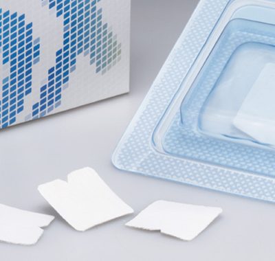
- BCG-MeRG (Biocollagen)
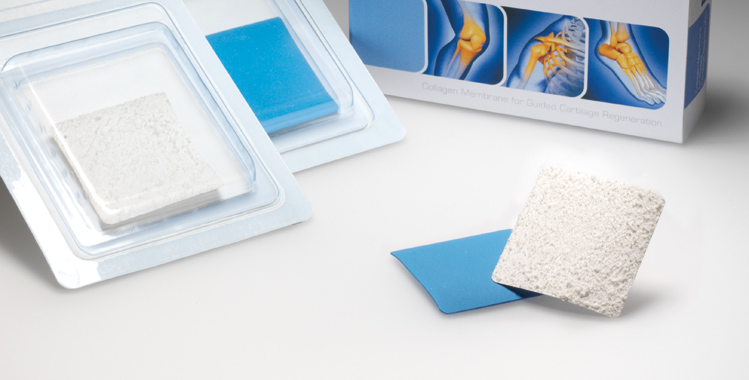
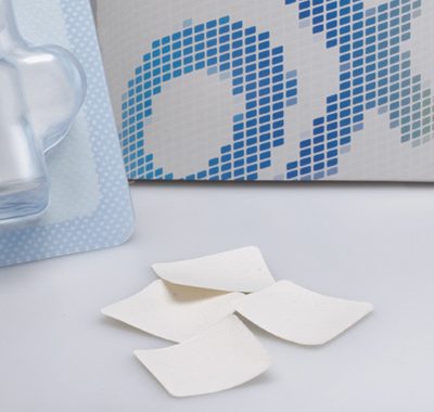
Collagen membrane
50 x 50 x 0.2 mm
Elastomer surgical template
50 x 50 x 0.2 mm - BCG-MeRG/S (Biocollagen)
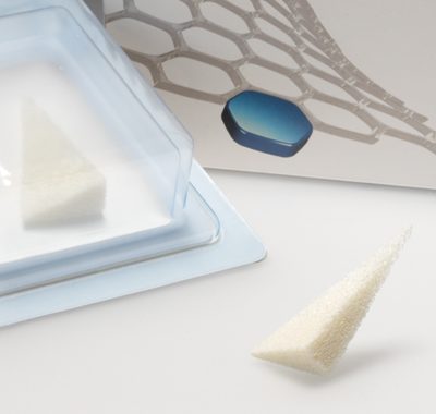
Collagen membrane
50 x 50 x 0.4 mm
Surgical template
50 x 50 x 0.2 mm - BCG-MeRG Q (Biocollagen)
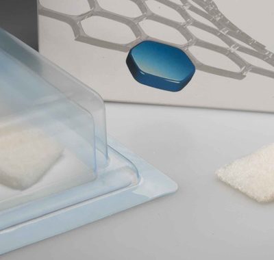
Collagen membrane
30 x 30 x 0.2 mm
Surgical template
30 x 30 x 0.2 mm - BCG-MeRG Disc (Biocollagen)
Collagen membrane
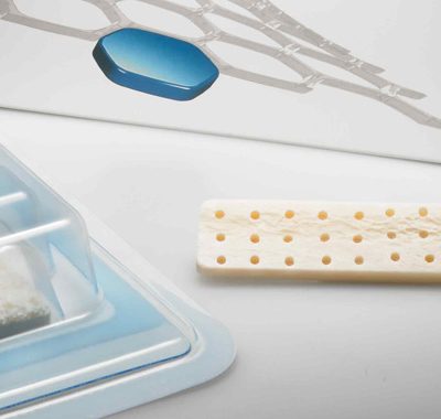
4 pieces: Ø 12-14-16-18 mm - BCG-MeRG K (Biocollagen)
Collagen membrane
30 x 30 x 0.2 mm
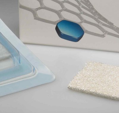
Non-friable, microfibrillar collagen membrane.
The specific layout of the fibres, granted by an exclusive production process, makes the membrane resistant to traction, twisting and pulling. The membrane has one smooth side and another rough fibrillar side, which allows it to be fixed with fibrin sealant (and stitches where appropriate). The membrane’s adhesive properties increase with contact with blood. The membrane collagen is obtained from equine Achilles’ tendon (one of the purest sources of type I collagen). This animal is also free from risk of B.S.E.
The chemical and structural characteristics of this substance are those of a glycoprotein able to interact with the receptors of thrombocytes and fibroblasts, to enable the XII and VIII factor, and to form the basic structures of connective tissue.
The interaction of the collagen with the thrombocytes is important for blood coagulation because the link with the platelet integrins determines the de-granulation of the platelets and the release of factors enabling the coagulation cascade with the formation of the fibrin net. The fibrin net holds elements of the particulates forming the coagulation, which subsequently set to work for the chemotactic factors that induce fibroblast migration.
MeRG carries out a ‘curtain effect’ action with mesenchymal cells, preventing them from dispersing in the joint cavity. MeRG is formed from collagen fibres laid out in a net that encourages cell surface adhesion. The three-dimensional structure of MeRG eases histological repair.
Biopsies in animal testing have shown that during the histological repair process, the fibroblasts anchor to the collagen fibrils, proliferate and orientate in order to reshape the damaged tissue.
The collagen therefore encourages tissue repair.


Reviews
There are no reviews yet.